|
Companies turn to
us to manufacture,
purify, protect
and package their
critical products |
 |
ELISApackaging.com
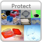
SK100, SK500 & SK900 Protective IVD Packaging
Benefits and Features
IVD packaging protects your diagnostics, gauges and reagents from damage. The high impact polypropylene container will not breakdown or discolor like chipboard. The packaging is also available with or without customized foam inserts.
Orders and Questions
Our on-line store can only accept orders shipped to a U.S. location. For international orders or for any questions regarding our products, services or store, please call Pozzetta.com sales at +1.303.783.3172 or email sales@pozzetta.com
|
|
| Specifications |
Part
Number |
Dimensions
(in/cm)
Description | Colors ** |
Price (USD) |
|
| |
|
|
|
|
| SK100 |
4 x 5 x 2.75 |
White, Black, |
$2.95 each |
|
| |
10.16 x 12.70 x 6.99 |
Blue, Pearl |
|
|
| SK500 |
6.5 x 7.75 x 4 |
White, Black, |
$4.50 each |
|
| |
16.51 x 19.69 x 10.16 |
Blue |
|
|
| SK900 |
8 x 12 x 4 |
White, Black |
$5.50 each |
|
| |
20.32 x 30.48 x 10.16 |
Blue |
|
|
| |
|
|
|
|
| Custom Foam Inserts |
|
|
|
SK100-Foam
|
Custom Foam Insert for SK100
case of 100 inserts |
Gray |
$195.00 case |
|
SK500-Foam
|
Custom Foam Insert for SK500
case of 100 inserts |
Gray |
$250.00 case |
|
SK900-Foam
|
Custom Foam Insert for SK900
case of 100 inserts |
Gray |
$350.00 case |
|
| |
|
|
|
|
| Stock Foam Inserts |
|
|
|
| SK100-151403 |
SK100 7 hole, Bottle, Blank |
Gray  |
$2.50 |
|
| SK100-130111 |
SK100 Foam Blank |
Gray |
$1.50 |
|
| SK100-150503 |
SK100-6 Hole |
Gray |
$1.50 |
|
| SK500-051209 |
SK500-Multi Holes + Microwell |
Gray |
$2.00 |
|
| SK500-032507 |
SK500-Mutiple Holes & Sizes |
Gray |
$2.00 |
|
| SK500-2013 |
SK500-Xtra Holes |
Gray |
$2.00 |
|
| SK900-2304 |
SK900-33 holes |
Gray |
$3.00 |
|
| |
|
|
|
|
|
|
| |
| Additional Products and Services |
Enzyme-linked immunosorbent assay, also called ELISA, enzyme immunoassay
or EIA, is a biochemical technique used mainly in immunology to
detect the presence of an antibody or an antigen in a sample. The
ELISA has been used as a diagnostic tool in medicine and plant pathology,
as well as a quality control check in various industries. In simple
terms, in ELISA an unknown amount of antigen is affixed to a surface,
and then a specific antibody is washed over the surface so that
it can bind to the antigen. This antibody is linked to an enzyme,
and in the final step a substance is added that the enzyme can convert
to some detectable signal. Thus in the case of fluorescence ELISA,
when light of the appropriate wavelength is shone upon the sample,
any antigen/antibody complexes will fluoresce so that the amount
of antigen in the sample can be inferred through the magnitude of
the fluorescence. Performing an ELISA involves at least one antibody
with specificity for a particular antigen. The sample with an unknown
amount of antigen is immobilized on a solid support (usually a polystyrene
microtiter plate) either non-specifically (via adsorption to the
surface) or specifically (via capture by another antibody specific
to the same antigen, in a "sandwich" ELISA). After the antigen is
immobilized the detection antibody is added, forming a complex with
the antigen. The detection antibody can be covalently linked to
an enzyme, or can itself be detected by a secondary antibody which
is linked to an enzyme through bioconjugation. Between each step
the plate is typically washed with a mild detergent solution to
remove any proteins or antibodies that are not specifically bound.
After the final wash step the plate is developed by adding an enzymatic
substrate to produce a visible signal, which indicates the quantity
of antigen in the sample. Traditional ELISA typically involves chromogenic
reporters and substrates which produce some kind of observable color
change to indicate the presence of antigen or analyte. Newer ELISA-like
techniques utilize fluorogenic, electrochemiluminescent, and real-time
PCR reporters to create quantifiable signals. These new reporters
can have various advantages including higher sensitivities and multiplexing[1][2].
Technically, newer assays of this type are not strictly ELISAs as
they are not "enzyme-linked" but are instead linked to some non-enzymatic
reporter. However, given that the general principles in these assays
are largely similar, they are often grouped in the same category
as ELISAs. Contents [hide] 1 Applications 2 History 3 Types 3.1
"Indirect" ELISA 3.2 Sandwich ELISA 3.3 Competitive ELISA 3.4 Reverse
ELISA 4 See also 5 References 6 External links [edit] Applications
ELISA results using S-OIV A neuraminidase antibody at 1 μg/ml to
probe the immunogenic and the corresponding seasonal influenza A
neuraminidase peptides at 50, 10, 2 and 0 ng/ml.Because the ELISA
can be performed to evaluate either the presence of antigen or the
presence of antibody in a sample, it is a useful tool for determining
serum antibody concentrations (such as with the HIV test[3] or West
Nile Virus). It has also found applications in the food industry
in detecting potential food allergens such as milk, peanuts, walnuts,
almonds, and eggs.[4] ELISA can also be used in toxicology as a
rapid presumptive screen for certain classes of drugs. The ELISA,
or the enzyme immunoassay (EIA), was the first screening test widely
used for HIV because of its high sensitivity. In an ELISA, a person's
serum is diluted 400-fold and applied to a plate to which HIV antigens
are attached. If antibodies to HIV are present in the serum, they
may bind to these HIV antigens. The plate is then washed to remove
all other components of the serum. A specially prepared "secondary
antibody" — an antibody that binds to other antibodies — is then
applied to the plate, followed by another wash. This secondary antibody
is chemically linked in advance to an enzyme. Thus, the plate will
contain enzyme in proportion to the amount of secondary antibody
bound to the plate. A substrate for the enzyme is applied, and catalysis
by the enzyme leads to a change in color or fluorescence. ELISA
results are reported as a number; the most controversial aspect
of this test is determining the "cut-off" point between a positive
and negative result. A cut-off point may be determined by comparing
it with a known standard. If an ELISA test is used for drug screening
at workplace, a cut-off concentration, 50 ng/mL, for example, is
established, and a sample will be prepared which contains the standard
concentration of analyte. Unknowns that generate a signal that is
stronger than the known sample are "positive". Those that generate
weaker signal are "negative." ELISA can also be used to determine
the level of antibodies in faecal content...specifically the direct
method [edit] History Before the development of the EIA/ELISA, the
only option for conducting an immunoassay was radioimmunoassay,
a technique using radioactively-labeled antigens or antibodies.
In radioimmunoassay, the radioactivity provides the signal which
indicates whether a specific antigen or antibody is present in the
sample. Radioimmunoassay was first described in a paper by Rosalyn
Sussman Yalow and Solomon Berson published in 1960[5]. Because radioactivity
poses a potential health threat, a safer alternative was sought.
A suitable alternative to radioimmunoassay would substitute a non-radioactive
signal in place of the radioactive signal. When enzymes (such as
peroxidase) react with appropriate substrates (such as ABTS or 3,3’,5,5’-Tetramethylbenzidine),
this causes a change in color, which is used as a signal. However,
the signal has to be associated with the presence of antibody or
antigen, which is why the enzyme has to be linked to an appropriate
antibody. This linking process was independently developed by Stratis
Avrameas and G.B. Pierce[6]. Since it is necessary to remove any
unbound antibody or antigen by washing, the antibody or antigen
has to be fixed to the surface of the container, i.e. the immunosorbent
has to be prepared. A technique to accomplish this was published
by Wide and Jerker Porath in 1966.[7] In 1971, Peter Perlmann and
Eva Engvall at Stockholm University in Sweden, and Anton Schuurs
and Bauke van Weemen in The Netherlands, independently published
papers which synthesized this knowledge into methods to perform
EIA/ELISA.[8][9] [edit] Types [edit] "Indirect" ELISA This article
may be confusing or unclear to readers. Please help clarify the
article; suggestions may be found on the talk page. (March 2009)
The steps of "indirect" ELISA follows the mechanism below: The antigen
to be tested for is added to each well of a microtiter plate, where
charges for many different conformations of proteins are present.
A solution of non-reacting protein, such as bovine serum albumin,
or casein is added to block any additional charges that did not
attract the protein of interest. Then the serum is added, which
contains antibodies of unknown concentration specific for the antigen
added originally. Afterwards, a secondary antibody is added, which
is specific for all antibodies from the species of the antibodies
added originally. This secondary antibody often has an enzyme attached
to it, which has no effect on the bonding properties of the molecule.
A substrate for this enzyme is then added. Often, this substrate
changes color upon reaction with the enzyme. The higher the concentration
of the enzyme that was present in the serum, the stronger the color
change. Often a spectrometer is used to give quantitative values
for color strength. The enzyme acts as an amplifier; even if only
few enzyme-linked antibodies remain bound, the enzyme molecules
will produce many signal molecules. A major disadvantage of the
indirect ELISA is that the method of antigen immobilization is non-specific;
any proteins in the sample will stick to the microtiter plate well,
so small concentrations of analyte in serum must compete with other
serum proteins when binding to the well surface. The sandwich ELISA
provides a solution to this problem. ELISA may be run in a qualitative
or quantitative format. Qualitative results provide a simple positive
or negative result for a sample. The cutoff between positive and
negative is determined by the analyst and may be statistical. Two
or three times the standard deviation is often used to distinguish
positive and negative samples. In quantitative ELISA, the optical
density or fluorescent units of the sample is interpolated into
a standard curve, which is typically a serial dilution of the target.
[edit] Sandwich ELISA A sandwich ELISA. (1) Plate is coated with
a capture antibody; (2) sample is added, and any antigen present
binds to capture antibody; (3) detecting antibody is added, and
binds to antigen; (4) enzyme-linked secondary antibody is added,
and binds to detecting antibody; (5) substrate is added, and is
converted by enzyme to detectable form.A less-common variant of
this technique, called "sandwich" ELISA, is used to detect sample
antigen. The steps are as follows: Prepare a surface to which a
known quantity of capture antibody is bound. Block any non specific
binding sites on the surface. Apply the antigen-containing sample
to the plate. Wash the plate, so that unbound antigen is removed.
Apply primary antibodies that bind specifically to the antigen.
Apply enzyme-linked secondary antibodies which are specific to the
primary antibodies. Wash the plate, so that the unbound antibody-enzyme
conjugates are removed. Apply a chemical which is converted by the
enzyme into a color or fluorescent or electrochemical signal. Measure
the absorbency or fluorescence or electrochemical signal (e.g.,
current) of the plate wells to determine the presence and quantity
of antigen. The image to the right includes the use of a secondary
antibody conjugated to an enzyme, though technically this is not
necessary if the primary antibody is conjugated to an enzyme. However,
use of a secondary-antibody conjugate avoids the expensive process
of creating enzyme-linked antibodies for every antigen one might
want to detect. By using an enzyme-linked antibody that binds the
Fc region of other antibodies, this same enzyme-linked antibody
can be used in a variety of situations. Without the first layer
of "capture" antibody, any proteins in the sample (including serum
proteins) may competitively adsorb to the plate surface, lowering
the quantity of antigen immobilized.. A descriptive animation of
the application of sandwich ELISA to home pregnancy testing can
be found here. [edit] Competitive ELISA A third use of ELISA is
through competitive binding. The steps for this ELISA are somewhat
different than the first two examples: Unlabeled antibody is incubated
in the presence of its antigen. These bound antibody/antigen complexes
are then added to an antigen coated well. The plate is washed, so
that unbound antibody is removed. (The more antigen in the sample,
the less antibody will be able to bind to the antigen in the well,
hence "competition.") The secondary antibody, specific to the primary
antibody is added. This second antibody is coupled to the enzyme.
A substrate is added, and remaining enzymes elicit a chromogenic
or fluorescent signal. For competitive ELISA, the higher the original
antigen concentration, the weaker the eventual signal. The major
advantage of a competitive ELISA is the ability to use crude or
impure samples and still selectively bind any antigen that may be
present. (Note that some competitive ELISA kits include enzyme-linked
antigen rather than enzyme-linked antibody. The labeled antigen
competes for primary antibody binding sites with your sample antigen
(unlabeled). The more antigen in the sample, the less labeled antigen
is retained in the well and the weaker the signal). [edit] Reverse
ELISA A new technique uses a solid phase made up of an immunosorbent
polystyrene rod with 4-12 protruding ogives. The entire device is
immersed in a test tube containing the collected sample and the
following steps (washing, incubation in conjugate and incubation
in chromogenous) are carried out by dipping the ogives in microwells
of standard microplates pre-filled with reagents. The advantages
of this technique are as follows: The ogives can each be sensitized
to a different reagent, allowing the simultaneous detection of different
antibodies and different antigens for multi-target assays; The sample
volume can be increased to improve the test sensitivity in clinical
(saliva, urine), food (bulk milk, pooled eggs) and environmental
(water) samples; One ogive is left unsensitized to measure the non-specific
reactions of the sample; The use of laboratory supplies for dispensing
sample aliquots, washing solution and reagents in microwells is
not required, facilitating ready-to-use lab-kits and on-site kits.